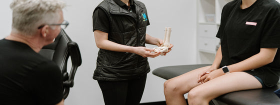
The shoulder joint is a ball and socket joint meaning it can move through large ranges of motion. The head of the humerus (ball) has a 4:1 ratio to the glenoid fossa, meaning only about 25% of the humeral head is in contact with the glenoid. Because of this, the shoulder joint is reliant on a large array of static stabilisers (glenoid labrum, joint capsule, glenohumeral ligaments) and dynamic stabilisers (rotator cuff muscles, biceps and triceps brachii tendon) to maintain stability.
What causes a shoulder dislocation?
The most common cause of a shoulder dislocation is a traumatic incident such as a fall onto an outstretched hand or a blow to the shoulder. Conditions that are susceptible to hypermobility such as Ehlers-Danlos can increase the risk of a dislocation as the joint has a larger degree of laxity.
Immediately post an anterior shoulder dislocation, severe pain which can radiate down the arm is the primary complaint. The first line of treatment is to reduce the shoulder dislocation. If this is not achieved immediately, it will be done at the hospital, usually after an X-Ray to rule out significant fractures and a Hill-Sachs lesion (dent in the head of the humerus).
Diagnosis:
Your physiotherapist will assess your shoulder function, stability and range through a range of orthopaedic tests. However, if the shoulder continues to feel weak and unstable, further imaging will be required even if the X-Ray is all clear.
Complications following shoulder dislocations include superior labrum anterior and posterior (SLAP) lesions, Bankart lesions (anterior labral tear) or tears to the glenohumeral ligaments (superior, middle and inferior). All these structures are difficult to review through an X-Ray alone.
So how do you know whether you need further imaging with an MRI?
1. Traumatic incident
2. Complaint of ongoing pain
3. Complaint of ongoing shoulder instability
4. Complaint of ongoing strength deficits
5. X-Ray cleared of fracture but the shoulder “does not feel right”
6. Positive shoulder instability test (apprehension test)
What can physiotherapy do?
Treatment for shoulder dislocations is highly dependant on the severity of the injury. A short period of immobilisation with a sling is needed to reduce pain and risk of further damage to the joint. After the initial phase of immobilisation, a structured shoulder strengthening rehabilitation program can be commenced to restore full shoulder range and function.
If the dislocation is more severe, a Bankart or SLAP lesions is present and the patient continues to report instability, a review with an Orthopaedic surgeon is recommended within the first 3-4 weeks post-injury (and yes our OHL Physio team can help link you up with a friendly shoulder-focused Orthopaedic surgeon in our network). If deemed appropriate by the surgeon and physiotherapist alike, an arthroscopic stabilisation procedure will be completed to provide stability to the glenohumeral joint.
Final thoughts:
Anterior shoulder dislocations are the most common types of shoulder dislocations and are often debilitating. It is important to seek appropriate medical attention to seek the right imaging, rehabilitation and referral pathways when appropriate. Our expert team of physiotherapist at the Optimal Health Lab are trained to rehabilitate shoulder dislocations and refer on to orthopaedic surgeons when appropriate. Book today if you’ve suffered such an event!!

OHL is integrating a new athletic screening assessment into its practice to further enhance our community's sporting ability. This screening assessment combines range of motion, strength profiling, force deck analysis, and subjective training status to give athletes a comprehensive performance snapshot. By establishing a baseline and identifying key areas for improvement, we can tailor your training to enhance performance, provide insight on key metrics, and stay resilient throughout the season. Whether you're preparing for preseason, managing midseason demands, or simply aiming to train smarter, this assessment delivers the data-driven insights you need.

If you're experiencing back or neck pain with neurological signs and symptoms, a thorough neurological examination is crucial for accurate assessment and effective treatment. In this Optimal Tip learn more about what we mean by completing a neurological exam!

Squats, deadlifts, and calf raises are key movement patterns that should be part of every strength and conditioning program—regardless of age and activity level. These functional movements support joint health, improve posture and balance, and reduce the risk of injury while building strength where it matters most.